Never proceed on your own without consultation.
The pathology report should document those histologic features important for guiding patient management, including those characteristics on which the diagnosis was based and also prognostic factors. Carcinoma refers to a malignant neoplasm of epithelial origin or cancer of the internal or external lining of the body. There was statistically significant difference between histological subtypes of melanoma and breslow thickness (p˂ 0.00 1). Spitzoid melanocytic neoplasms are named in honour of sophie spitz, an american pathologist, who initially proposed the term 'melanoma of childhood' The rarity of these tumors makes their identification and treatment a dilemma.

Ki67 antigen and pcna proliferation markers predict survival in anorectal malignant melanoma.
Non neoplastic pathology of the inner ear rarely requires biopsy. Tumor thickness and prognosis in clinical stage 1 malignant melanoma. The rarity of these tumors makes their identification and treatment a dilemma. It has been suggested they are distinct from teratomas. Causes second most common type of skin cancer epidermal keratinocytes acquire antiapoptotic properties → frequent mitosis risk factors immunosuppression, chronic uv exposure, fair skin, albinism, xeroderma. Other aspects of melanoma, including the pathologic approach to evaluation of. Incidence over 300,000 new cases skin tumors every year in usa. If this creates a problem for you, please contact us by completing the form below. Chapter 5 malignant skin tumors figure 5.5 the gross pathology of the heart in a case of metastatic melanoma. B16 melanoma, pp2a, connexin, nop5/sik family, subtracted cdna library doi: Due to lack of capacity, we unfortunately have to shut down oncolex.org. Epithelial tissue is found throughout the body. There was also midthoracic pain.
Moreover, similar roles were expected for the genes in the process by which human melanoma cells metastasize. Pathologists often practice as consultant physicians who develop and apply their knowledge of tissue and laboratory analyses to assist in the. Causes second most common type of skin cancer epidermal keratinocytes acquire antiapoptotic properties → frequent mitosis risk factors immunosuppression, chronic uv exposure, fair skin, albinism, xeroderma. Ewing's sarcoma is a malignant, distinctive small round cell sarcoma associated with a t (11:22) translocation and most commonly occurs in the diaphysis of long bones. A surgical pathology vade mecum.

Closed loops and networks matrix patterns are seen.
Due to lack of capacity, we unfortunately have to shut down oncolex.org. Pathologic examination of sentinel lymph nodes provides very important prognostic information. Nail melanoma can be obvious, but can hide in brown and even red bands, and can be amelanotic, dr. There was also midthoracic pain. Ki67 antigen and pcna proliferation markers predict survival in anorectal malignant melanoma. Other features of benignity are confinement within investing sclerosis and, if previously biopsied, association with hemorrhage and/or fibrotic biopsy tract. It is composed of spindle and epithelioid cells and has up to 15 mitotic figures per 10 hpf. Analysis of 3661 patients from a single center. The accuracy of any histopathology report is at least partly dependent on the amount of tissue provided and the availability of relevant clinical details. (3 x 5 = 15) 1. Closed loops and networks matrix patterns are seen. A revision of the 1972 sydney classification. Symptoms include dizziness, hearing loss and tinnitus.
Diagnosis is made with a biopsy showing sheets of monotonous small round blue. Metastatic adenocarcinoma of lung (a), follicular thyroid carcinoma (b), malignant melanoma (low power) (c), squamous cell carcinoma (d), clear cell renal carcinoma (e), and melanoma (high power) (f). Spitzoid melanocytic neoplasms are named in honour of sophie spitz, an american pathologist, who initially proposed the term 'melanoma of childhood' A revision of the 1972 sydney classification. Statistical categorization of human histological images.

Analysis of 3661 patients from a single center.
Invasive melanoma histopathology reporting guide. Distribution of head and neck lesions diagnosed on histopathology in…. 9000 are melanomas, that is. Closed loops and networks matrix patterns are seen. Note the fingerlike projections of the tumor. For all surgically enucleated eyes, call the attending pathologist to determine the best way to work up the case. In the present study, 53 omms (43.1%) were detected with macular lesions and 56.9% of patients (n = 70) had nodular melanomas. Giant cell rich histology can be seen in both benign and malignant tumors and can be found in diverse sites from the skeleton to the head and neck to the viscera. Moreover, similar roles were expected for the genes in the process by which human melanoma cells metastasize. Return to the general pathology menu. It covers diagnostic surgical pathology, cytology, autopsy practice, histological technique, lab management, rcpath guidance and uk law relevant to histopathology. Vascular changes in diabetic retinopathy. Chapter 5 malignant skin tumors figure 5.5 the gross pathology of the heart in a case of metastatic melanoma.
39+ Histopathology Malignant Melanoma Histology Diagram PNG. 9000 are melanomas, that is. It has been suggested they are distinct from teratomas. The most frequent subtype for melanoma based on histopathology was the acral lentiginous melanoma (alm) (74 patients, 38.3%). Nail melanoma can be obvious, but can hide in brown and even red bands, and can be amelanotic, dr. 90% of schwannomas are solitary and sporadic.
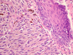
A key feature that helps to establish the diagnosis of benign, entrapped glands is background benign histology (ie, there is no indication of a neoplastic process in the idp or elsewhere) malignant melanoma histology. Causes second most common type of skin cancer epidermal keratinocytes acquire antiapoptotic properties → frequent mitosis risk factors immunosuppression, chronic uv exposure, fair skin, albinism, xeroderma.